


 |

Information about the heart, coronary artery disease, cardiac
catherization, treatment for
cardiac catherization, balloon angioplasty,
and coronary stent
implantation.
|
The Heart |
|
The heart is a muscle located in your chest under the lower
part of your sternum (breast bone). In healthy adults, the heart is usually about
the size of a man's closed fist. The job of the heart is to pump blood to all parts
of the body by way of blood vessels (elastic tubes) which are called arteries and
veins. Arteries carry blood filled with oxygen and other nutrients to the body; veins
carry "used" blood from the body back to the heart. The heart also sends
the blood to the lungs where the oxygen we breathe is added to the blood.
The heart is divided into right and left sides, and upper and lower chambers. The
upper chambers, called atria, receive blood and pass it on to the lower chambers, called
the ventricles. The ventricles pump blood out of the heart, the right ventricle to
the lungs, and the left ventricle to the body. Blood flow into and out of the
chambers of the heart is controlled by valves. These valves open and close like
doors to keep the blood moving in one direction.
Blood flow through the heart, lungs, and the body is called circulation. As the
heart pumps, it produces a pulse. Counting the pulse tells us how many times the
heart beats in one minute.
In order for the heart to be able to do its work, it must be fed with oxygen-filled
blood. The blood vessels which feed the heart muscles are called coronary arteries.
The heart pumps the
blood through the circulatory system and
throughout the body. There are four
valves within the heart. Any may have
problems requiring medical attention.
Return to
Top
|
|
Coronary Artery Disease |
|
Having Coronary Artery
Disease (CAD) means that the inside of one or
more of your coronary arteries has become
narrowed, or blocked with fatty deposits,
calcium, or other materials. This is the
most common problem found in the heart.
Because of this narrowing,
part of your heart does not always get enough
oxygen-filled blood to do its work. This
usually causes chest discomfort, shortness of
breath, pressure, or pain. If you have
experienced any of these sensations, your doctor
has probably referred to this discomfort as
angina.
Risk factors for Coronary
Artery Disease include: smoking, high
blood pressure, high levels of fatty substances
in blood, excessive weight, lack of exercise,
and stress. Other risk factors are
diabetes mellitus and a family history of heart
disease.
It is very important for
patients with CAD to know the risk factors so
they can do something to decrease the risk of
their disease getting worse. This includes
eliminating or modifying the risk factors that
you have control over. Your doctor may
suggest ways to lose weight, reduce stress, or
increase exercise.
Other heart problems,
different from CAD, can occur in the valves or
in the heart muscle itself. One way
doctors diagnose CAD or other heart problems is
through cardiac catheterization.
Return to
Top
|
|
Cardiac Catheterization |
|
A
cardiac catheterization is a special x-ray study
of the heart. A flexible narrow plastic
tube called a catheter is inserted into a vein
or an artery in your arm or upper leg. The
catheter is then passed through the blood vessel
into a heart chamber or to a coronary
artery. Special x-ray dye, called
contrast, is injected and allows your doctor to
see the chambers, valves, or coronary arteries
in your heart on an x-ray television
screen. Certain pressures within the heart
may be measured and blood samples may be drawn.
Your heart has three main
coronary arteries. On the left, the Left
Main coronary splits into the Left Anterior
Descending that is on the front of the heart and
the Left Circumflex artery that travels around
the left side and to the back. The third
is the Right Coronary Artery that wraps around
the right and bottom of the heart. Each
artery is responsible for blood flow to a
certain section of heart muscle.
Once your doctor feels a
cardiac cath should be performed, he/she will
talk to you about it and try to decide on the
best time to have it done.
Return to
Top
|
|
Treatment for Coronary Artery Disease |
|
Following
your cardiac catherization, your doctor will
review the x-ray films taken during the
procedure and determine if there is narrowing or
blockage requiring further treatment. Your
doctor will discuss the results of your
angiogram with you and advise treatment options
based on your medical history, individual
anatomy, and personal needs. The treatment
options available may include medication,
balloon angioplasty or coronary artery bypass
graft surgery. If angioplasty is the best
treatment for your coronary artery disease, it
may also include the placement of a coronary
stent.
Return to
Top
|
|
Balloon Angioplasty |
|
The
setup and preparation for the balloon
angioplasty procedure is very similar to a
cardiac catherization procedure. The
difference in balloon angioplasty, also known as
Percutaneous Transluminal Coronary Angioplasty
or PTCA, is that a small balloon attached to a
catheter is passed into the artery to the site
of the blockage or lesion. The balloon is
then inflated to compress the fatty plaque
deposits and expand the artery in order to
increase blood flow through the artery.
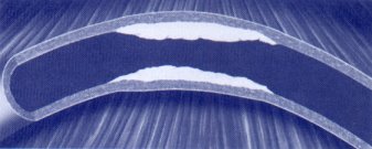 Pre-Angioplasty
Narrowing Pre-Angioplasty
Narrowing You will have an intravenous (IV) infusion of
the medication heparin which is an anticoagulant
that works as a blood thinner to aid in the
prevention of blood clot formation or
thrombus. If you feel discomfort at any
time during the PTCA procedure you are
encouraged to tell the doctor or nursing staff
about it immediately.
 Post-Angioplasty Enlarged
Lumen Post-Angioplasty Enlarged
Lumen Although approximately
400,000 balloon angioplasty procedures are
performed each year in the United States,
studies have shown that 30-50% of those patients
develop a re-narrowing or restenosis of the
lesion within 3-12 months after the
procedure. If your doctor feels it is
necessary, he or she may recommend placement of
a coronary stent, which is a device designed to
keep the treated area of the artery open after
PCTA.
Return to
Top
|
|
Coronary Stent
Implantation |
|
A coronary stent is a device made of medical grade stainless steel or some
other type of metal. It is shaped like a small tube that acts as a scaffold against
the artery wall. The stent is mounted on a balloon catheter. 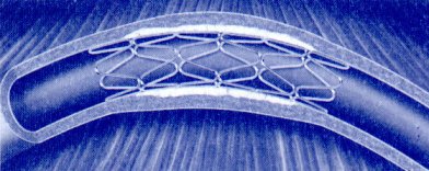
Balloon
Withdrawn-Stent Implanted
The stent and balloon
are advanced to the angioplasty site, the balloon is inflated, and the stent expands
against the inner wall of the artery. It may be necessary to re-inflate the balloon
multiple times to fully expand the stent. Your doctor may also decide to implant
more than one stent to adequately cover the diseased area and minimize the chance of
restenosis or abrupt closure of the artery.
After the stent is fully expanded, the balloon is removed and the stent remains
permanently implanted to hold the artery open, improving blood flow and relieving symptoms
of angina or chest pain.
Within several weeks
after the stent implantation, the body will grow
tissue over the stent ensuring that the device
will remain in its intended position in the
artery.
Return to
Top
|
Vijay M. Haryani, M.D., FACC
455 West Court Street, Suite 302, Kankakee, Illinois 60901
(Phone): 815-936-3240 * (FAX): 815-936-3243
(Email): info@haryani.com
|
|
